OUR SERVICES
Latest Technology in Eyecare
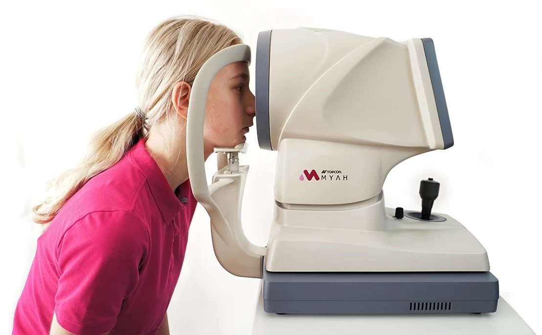
Axial Length Measurement for Children: Accurate Myopia Tracking with Topcon MYAH
We include axial length measurement on all children’s eye exams using the Topcon MYAH which is a dedicated, state-of-the-art instrument for axial length assessment and pediatric myopia management.
Why axial length matters
Axial length (the front-to-back length of the eye) is the most reliable indicator of long-term myopia progression. Tracking axial length growth provides objective data that predicts future refractive error and the risk of high myopia.
Measuring axial length is more accurate than relying on glasses prescriptions alone, especially in growing children whose refractive error can change rapidly.
How axial length measurement helps your child
Monitor growth: Measure and track axial length over time to detect early progression.
Predict refractive changes: Use axial length trends to estimate future nearsightedness risk.
Evaluate treatment effectiveness: Objectively assess whether myopia control strategies are working.
Personalize treatment: Tailor myopia management (orthokeratology, low‑dose atropine, specialty contact lenses) based on biometric data, corneal shape, and axial length trends.
When your optometrist will perform axial length measurement
On all pediatric exams at our clinic
When assessing a child with early or progressing myopia
Before and during myopia management to monitor treatment response
Why we use MYAH in our myopia management program because axial length is the single most important metric for long-term myopia control, the Topcon MYAH is central to our evidence-based approach. Its precise biometric and corneal measurements allow our team to design personalized myopia control plans and provide clear, objective progress reports to families.
Book a pediatric eye exam to include axial length measurement and learn whether myopia management is recommended for your child.
VISUAL FIELD TESTING

Our optometrists may recommend you to have your peripheral vision checked. At View Optometry we can do this by using an instrument called a visual field. You may also require a visual field test for your driver’s license. This additional testing would be fully covered by your B.C. Care Card.
Visual field testing is essential for detecting and monitoring:
Glaucoma
Optic nerve damage
Neurological conditions (stroke, pituitary tumors, brain lesions)
Medication-related vision changes
This test is quick, non-invasive, and critical for identifying blind spots or visual pathway issues that cannot be detected during a routine eye exam.
Reasons the optometrist may want you to perform a visual field test:
If the doctor suspects you may be developing glaucoma
If you have a family history of glaucoma
If you had or the doctor suspects you have had a stroke or tumor in the brain
If you have an ocular disease that affects the optic nerve
To determine if a lid droop is affecting your vision
To obtain to driver’s license
Baseline testing
Ocular Coherence Tomography (OCT)
What is an OCT?
An OCT uses light waves to take a scan of the back or front of your eye. It is similar to an ultrasound image in that it takes a cross section image of a part of your body with non-invasive techniques. Every eye exam at our clinic includes an OCT scan of your eyes.
This technology helps diagnose and manage:
Glaucoma
Macular degeneration
Diabetic retinopathy
Retinal swelling and structural abnormalities
The OCT also provides 3D imaging, RNFL (retinal nerve fiber layer) analysis, ganglion cell layer measurements, and progression reports, offering unparalleled insight into ocular health.
Reasons the optometrist may want to take an OCT scan of your eyes:
If the doctor suspects you have a retinal disorder, glaucoma, or a corneal disorder
To monitor progression of an eye disease
If you have diabetes
If you have a family history of macular degeneration
Baseline testing

Meibography: What it is and why we use it
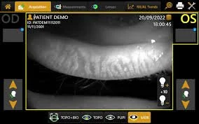
Meibography is a specialized imaging test that visualizes the meibomian glands inside your eyelids. At View Optometry we use the Topcon MYAH to capture clear images of these glands and assess their health.
Why meibomian glands matter
Meibomian glands produce meibum, the oily outer layer of the tear film.
Healthy meibum prevents rapid tear evaporation and keeps eyes comfortable.
When these glands are blocked, damaged, or reduced, patients commonly experience dry, irritated eyes and a higher risk of styes.
When your optometrist recommends meibography
To check for meibomian gland blockage or loss.
To monitor whether a prescribed treatment (warm compresses, lid hygiene, in-office procedures, or medications) is improving gland function.
As a visual tool to show patients the condition of their glands and explain treatment options.
Conditions diagnosed and managed with meibography
Meibomian gland dysfunction (MGD)
Evaporative dry eye disease
Blepharitis and other eyelid inflammatory conditions
Ocular surface disease
Contact lens–related dry eye
Benefits of meibography with Topcon Myah
Noninvasive, quick imaging for accurate gland assessment
Helps guide targeted treatment plans for dry eye and eyelid disease
Provides a baseline and follow-up images to track progress
If you have persistent dryness, burning, fluctuating vision, or recurrent styes, ask our North Vancouver optometrists about meibography, it can reveal hidden causes and help us create an effective treatment plan.
Cornea Topography
Corneal topography is like a map of the surface of your cornea, the clear front part of your eye. It shows how curved or flat different areas of the cornea are.
Reasons your optometrist would do topography:

To screen for a cornea disease called keratoconus which is common in patients with high astigmatism
To perform specialty contact lens fittings
Often needed prior to refractive surgery to determine if you are a good candidate for the surgery
Our investment in advanced diagnostic technology delivers faster, more accurate eye care that protects vision and improves outcomes. Benefits include:
Earlier detection of eye disease, allowing timely treatment and better prognosis
More accurate diagnoses through high-resolution imaging and objective testing
Precise biometric measurements for improved cataract, LASIK and contact lens planning
Highly effective myopia management with data-driven progression tracking
Objective dry eye evaluation to guide targeted therapies and relief
Better long-term monitoring to catch subtle changes over time
More effective, customized treatment plans based on comprehensive diagnostic data
A more comfortable, efficient exam experience with streamlined testing workflows
By combining cutting-edge technology with personalized care, we safeguard your vision, optimize treatment results, and enhance overall eye health.